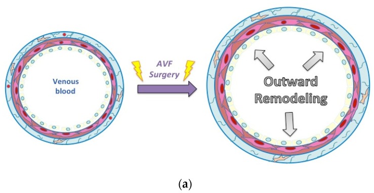
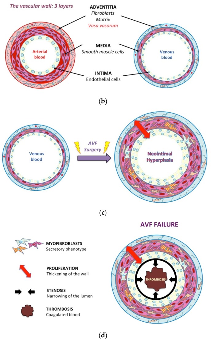
Figure 2.
Mechanisms of venous wall remodeling. (a) Similar to the arterial wall, the venous wall is composed of three layers: the external layer, called the adventitia, composed of extracellular matrix (ECM), fibroblasts, immune cells, and vasa vasorum. The middle layer, called the media, is made of smooth muscle cells (SMC) and some extracellular fibers. The internal layer, also called intima, is composed of endothelial cells. (b) and (c), upon arteriovenous fistula (AVF) creation, in response to the altered hemodynamic environment, several structural changes occur in the vessel wall. Surgery induces endothelial denudation, matrix reorganization to accommodate outward expansion but also vascular SMC proliferation and migration, contributing to NH formation. (d) Aggressive intimal hyperplasia induces stenosis (narrowing) of the vessel that can provoke thrombosis (occlusion).


