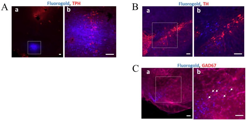
Figure 4.
Retrograde labeling of neurons following fluorogold (FG) injection into the median raphe nucleus (MnRN). (A) The square on the confocal laser-scanning microscope images under low (a) and high (b) magnification indicates the MnRN region at 3 days after FG injection. The image shows that FG (blue) was precisely injected into the MnRN. Tryptophan hydroxylase (TPH)-positive cells (red) were observed in the MnRN. Scale bar: 100 µm. (B) and (C) The square on the photomicrograph taken under low (a) and high (b) magnification indicates the substantia nigra pars compacta (SNpc) (B) and the reticular part of the reticular part of the substantia nigra (SNr) (C), which were analyzed using a confocal laser-scanning microscope at 3 days after FG injection. Scale bar: 100 µm. FG-labeled cells (blue) (B-a and -b, C-a and -b) and GAD67-positive cells (red) (C-a and -b) were co-localized in the SNr regions (indicated by white arrows) (C-b), but TH-positive cells (red) (B-a and -b) were not co-localized in the SNpc regions.

