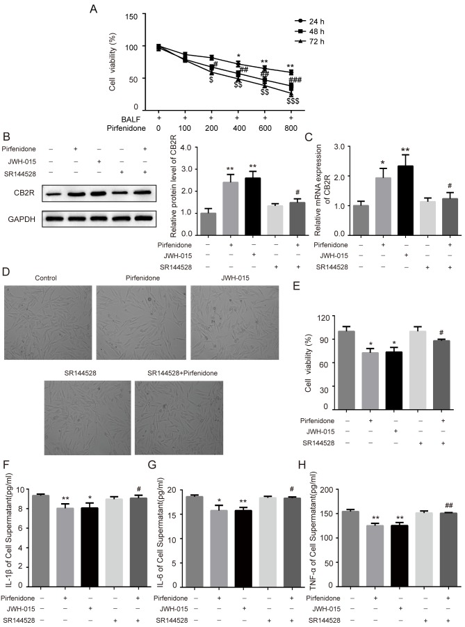
Figure 4.
Activation of CB2R mediates the protective effect of PFD on BALF-treated WI38 cells. WI38 cells were incubated with BALF from bleomycin-treated mice and treated with 200 µg/ml pirfenidone, 20 µM JWH-015, 1 µM SR144528, and 200 µg/ml pirfenidone + 1 µM SR144528. (A) The MTT assay was performed to determine the viability of WI38 cells treated with various concentrations of pirfenidone (0, 100, 200, 400, 600, and 800 µg/ml) for 24, 48 or 72 h. Data are presented as the mean ± SEM. *P<0.05, **P<0.01 vs. cell group only treated with BALF for 24 h; #P<0.05, ##P<0.01, ###P<0.001 vs. cell group only treated with BALF for 48 h; $P<0.05, $$P<0.01, $$$P<0.001 vs. cell group only treated with BALF for 72 h. The protein and mRNA levels of CB2R were detected by (B) western blotting and (C) RT-qPCR. (D) WI38 cell proliferation was observed under a light microscope (magnification, ×40) and detected by MTT assay. (E) Quantitative analysis of cell proliferation rate. The levels of (F) IL-1β, (G) IL-6 and (H) TNF-α in cell culture supernatant were detected by ELISA. Data are presented as the mean ± SEM. *P<0.05, **P<0.01 vs. untreated control group; #P<0.05, ##P<0.01 vs. PFD only group. PFD, pirfenidone; CB2R, CB2 receptors; BALF, bronchoalveolar lavage fluid.

