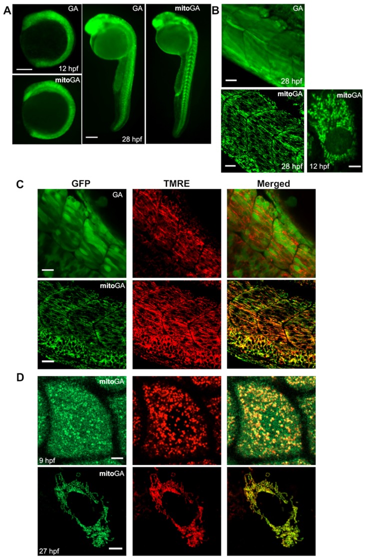
Figure 1.
Expression of biosensors GFP-aequorin (GA) and mitoGA in zebrafish embryos. (A) Ubiquitous transient expression of GA and mitoGA in zebrafish at 12 and 28 hpf visualized with a fluorescence stereomicroscope. (B) Localization of GA and mitoGA expressed in the trunk musculature by confocal microscopy (28 hpf) and mitoGA in the enveloping layer (12 hpf). (C) MitoGA, but not GA, colocalizes with the mitochondrial marker TMRE in skeletal muscle at 28 hpf. (D) Colocalization of mitoGA with TMRE at 9 and 27 hpf in the cells of the enveloping layer of a zebrafish embryo. Mitochondrial import of mitoGA was complete at 27 hpf. The scale bars represent 200 µm (A), 20 µm (B,C) and 5 µm (D).

