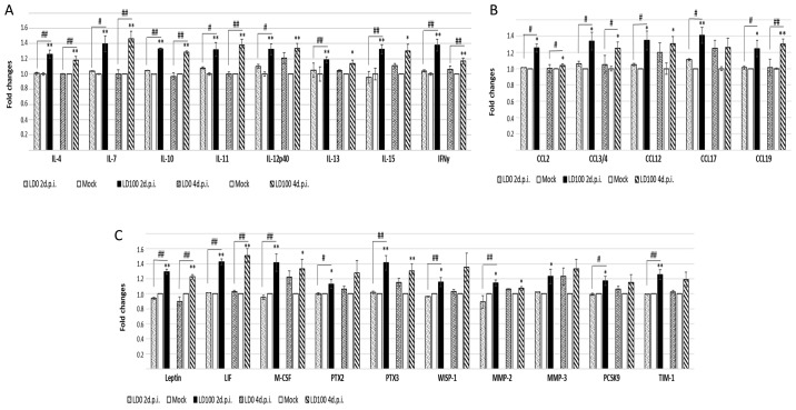
Figure 5.
Cytokines exhibiting increased expression in the lungs of the mice infected with a lethal dose of H1N1. BALB/c mice were infected intranasally with LD0 and LD100 doses of H1N1 virus. Protein expression levels of (A) interleukins, (B) chemokines and (C) proteins expressed during tissue damage or injury, were determined in lungs harvested at 2 and 4 days p.i. The values represent the mean of two separate experiments. *P<0.05, and **P<0.001 vs. mock. #P<0.05 and ##P<0.001, as indicated. H1N1, A/PR/8/34 virus; LD0, 101 plaque-forming units; LD100, 103 plaque-forming units; p.i., post-infection.

