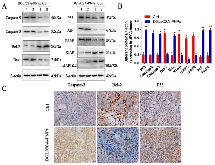
Figure 7.
Western blot and immunocytochemistry analysis of DGL/CSA-PNPs induced apoptosis. (A,B) extracts of JEG3 tumor from DGL/CSA-PNPs-administrated mice were used for anticancer mechanism analysis by Western blotting; (C) JEG3-tumor slices collected from DGL/CSA-PNPs-administrated mice were used for immunocytochemistry analysis of caspase-3, bcl-2, and P53 in vivo. Scale bar = 20 μm. *P < 0.05 and **P < 0.01 compared to untreated controls. The data were analyzed by using one-way ANOVA and presented as mean ± standard deviation (n = 2).

