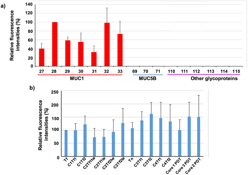
Figure 4.
Representative results of MUC1 glycopeptide microarray screening of antisera from Qβ-MUC1-Tf 17 immunized mice, (a) Comparison of fluorescence intensities of microarray components containing MUC1 glycopeptides bearing Tf antigen at various locations showed that glycosylation at that PDT*R region led to the strongest recognition by postimmune sera. Glycopeptide 27: PAHGVT*SAPDTRPAPGSTA; 28: PAHGVTSAPDT*RPAPGSTA; 29: PAHGVTSAPDTRPAPGST*A; 30: PAHGVT*SAPDT*RPAPGSTA; 31: PAHGVT*SAPDTRPAPGST*A 32: PAHGVTSAPDT*RPAPGST*A; 33: PAHGVT*SAPDT*RPAPGST*A. Glycopeptides 69–71 are various MUC5B glycopeptides. 110–115 are poly(LacNAc)-BSA, fetuin, transferrin, ICAM-1, porcine stomach mucin, and bovine submaxillary mucin, respectively, (b) Comparison of fluorescence intensities of microarray components containing MUC1 glycopeptides bearing various glycans at PAHGVTSAPDT*RPAPGSTA showed that, although Tf gave the strongest recognition, other glycans can be recognized, as well. Glycan structures: glycopeptide 28: Tf (for abbreviations and structures, see Supporting Information Scheme S2 and Figure S8); 35: C1Tf1; 42: C1Tf2; 56: C2Tf1he; 49: C2Tf1te; 73: C2Tf2he; 63: C2Tf2te; 21: Tn; 80: C3Tf1 87: C3Tf2; 94: C4Tf1; 101: C4Tf2; 109: core 1 PDT; 108: core 3 PDT; 107: core 2 PDT. The error bars represent standard deviation (SD) of eight replicates.

