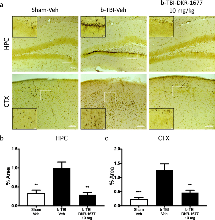
Figure 3.
Treatment with DKR-1677 (10 mg/kg/day) protects mice from neurodegeneration after Blast-Mediated TBI (b-TBI). Silver staining was used to determine neurodegeneration. (a) Representative pictures of the silver stained hippocampus (HPC) and cortex (CTX) of the sham-veh, b-TBI-veh, and b-TBI-DKR-1677 (10 mg/kg/day) groups. Silver staining quantification was performed measuring the % area covered by the staining (b and c). Scale bar = 100 μm in larger box and 10 μM in smaller box, as shown. Higher magnified inset is of the white-outlined box in the lower magnification image. Values are presented as mean ± SEM. Significance was determined by one-way ANOVA with multiple comparisons (n = 5/group). **p < 0.01, *** p < 0.001.

