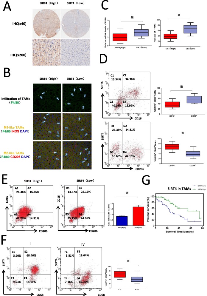
Fig. 2.
Downregulation of SIRT4 is associated with increased macrophage infiltration and M2 macrophages in HCC peritumour tissues. a Immunohistochemical staining was utilized to examine SIRT4 in HCC peritumour tissues (magnification at × 40 and × 200). b The cell phenotype in tissue sections from HCC patients was detected by F4/80+/iNOS+ for M1 and F4/80+/CD206+ for M2 using double immunohistofluorescence. The M1-like or M2-like macrophages are indicated by an arrow (400×). c QRT-PCR was used to detect F4/80 mRNA levels. Statistical analysis of the relationship between TAM infiltration and SIRT4 level. d FCM showed SIRT4 expression in CD206+ or CD206− TAMs and CD16+ or CD16− TAMs. e FCM analysis demonstrated TAM polarization in HCC peritumour tissues of different SIRT4 expression profiles. Statistical analysis between the ratio of M2/M1 macrophages and the SIRT4 level. f FCM analysis demonstrated SIRT4 expression in CD68+ TAMs from tumours with different grades. g. The Kaplan–Meier survival curve demonstrated the correlation between SIRT4 expression in CD68+ macrophages and survival of HCC patients. Data are displayed as the means ± SE, * p < 0.05

