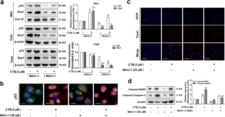
Fig. 7.
Activation of Drp1 is required for p53-dependent apoptosis a Mitochondrial translocation of Drp1 and p53 were detected by Western Blot. b Representative Fluorescence microscope imaging of SMMC-7721 cells labeled with DAPI, anti-p53 antibody and Mito-tracker Green. Scale bar: 20 μm. c TUNEL staining evaluated cells apoptosis. Red fluorescence indicated apoptotic cells. Scale bar: 50 μm. d Western blot analysis of cleaved-caspase-3 and cleaved-PARP levels in cytoplasm. Data are represented as mean ± SD. Significance: *P < 0.05, **P < 0.01 and ***P < 0.001 vs Control; #P < 0.05, ##P < 0.01 and ###P < 0.01 vs CTB (2 μΜ) treatment

