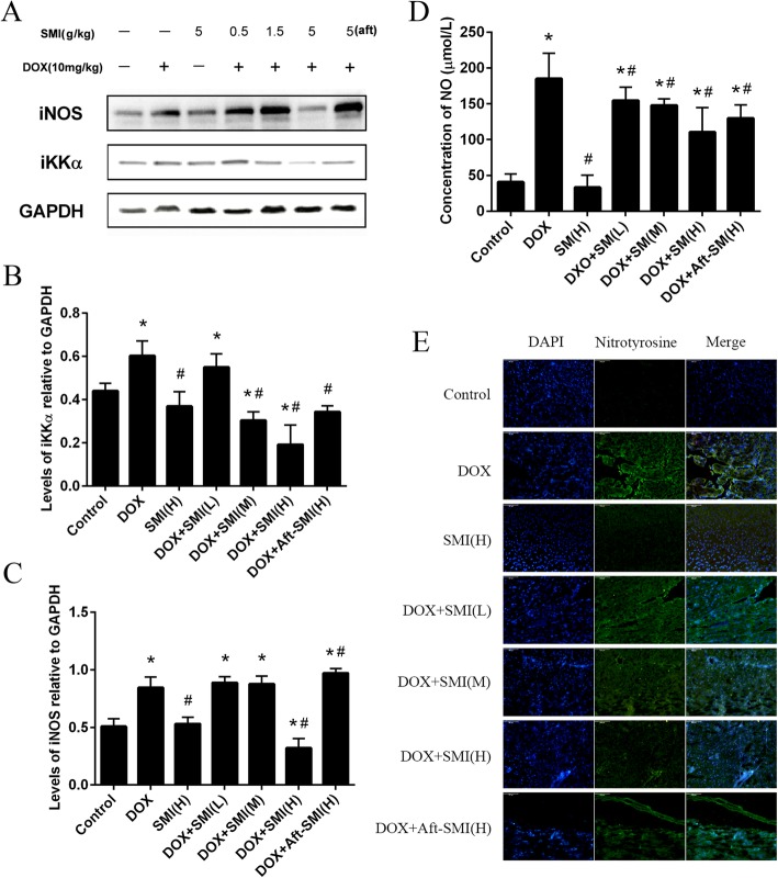
Fig. 7.
Mice received different treatment in experiment. And IKK-α and iNOS expressions were detected by Western blot analysis into tissue homogenates from mice heart; GAPDH protein expression was used as loading control (a – c). Effect of DOX on NO release was evaluated by NO level in the serum of mice (d). (Means ± S.D., n = 10). *, significantly different (P < 0.05) from respective values in the control group;#, significantly different (P < 0.05) from respective values in the DOX group. Nitrotyrosine production in heart of ICR mice treat with DOX or SMI (e). Frozen myocardial tissue sections were stained with Anti-Nitrotyrosine (green) and nucleus with DAPI (blue) and were determined by immunofluorescence analysis. (Bar 200 μm)

