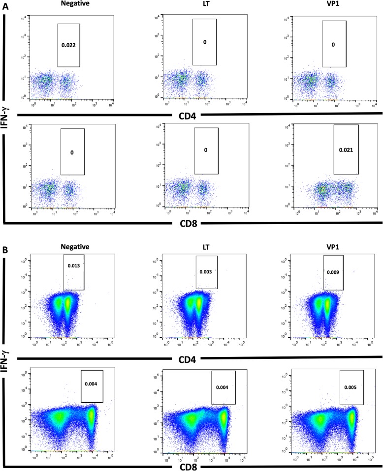
Fig. 3.
BKPyV-specific CD4+ (upper panel) and CD8+ (lower panel) T cells with IFN-γ production in KT recipient; patient number 8. Peripheral blood mononuclear cells were tested by intracellular cytokine staining for production of IFN-γ after stimulation with LT and VP1 antigens at diagnosis (a) and after adjustment of immunosuppression (b). These IFN-γ-producing cells were determined on the basis of their forward and side scatter plots

