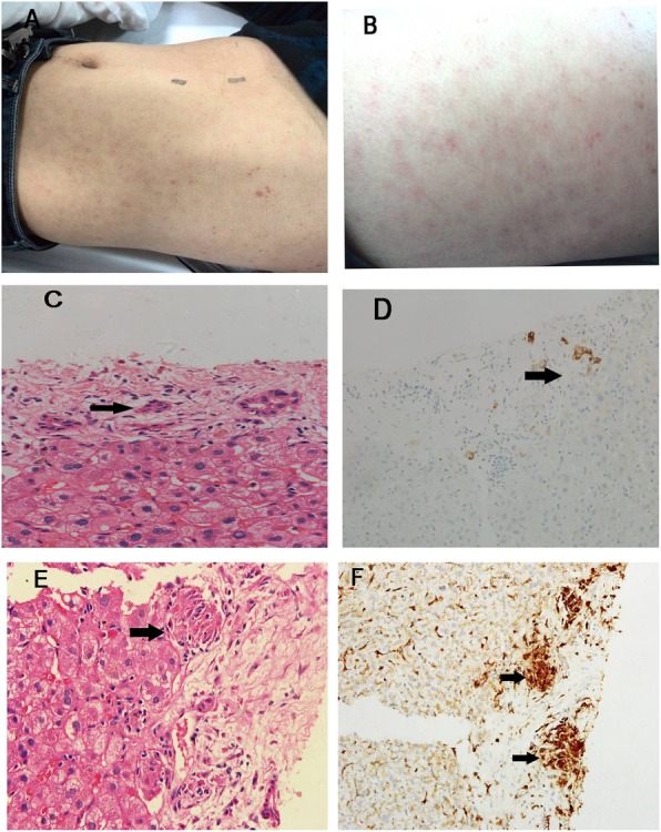
Fig. 1.

Patient’s body appearance and histological findings. a multiple erythematous macules and hyperpigmentation on the back; b, multiple erythematous macules and hyperpigmentation over the belly; c, HE staining shows bile duct epithelial cell injury, atrophy cholangiocyte, and portal tract inflammation (× 400); d, CK7 staining of cholangiocyte reveals atrophy cholangiocyte and bile duct lesion (× 100); e, HE staining shows granulomas (× 400); f, CD68 staining of macrophagocyte shows granulomas (× 100). Black arrows indicate lesions
