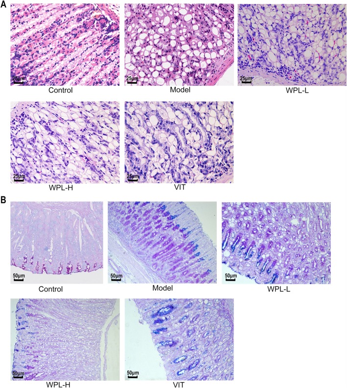
Fig. 2.
Histological evaluation of gastric epithelial dysplasia and intestinal metaplasia (H&E staining, 400×). a The model gastric epithelium displayed dysplasia pathology characterized by glandular architectural abnormalities. After WPL intervention, these dysplasia pathological alterations, especially irregularities of glandular structure, were regressed to varying degrees. n = 6 in each group. b Histological evaluation of gastric intestinal metaplasia (AB-PAS staining, 200×). Neutral mucins present in normal mucosa were stained red. Sialomucins expressed only in intestinal-type metaplasia (IM) were stained blue. Images of the model gastric epithelium depicted prominent IM lesions, which were dramatically reduced after WPL administration. n = 5 in each group

