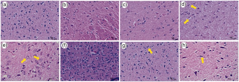
Figure 4.
Histological changes in hypothalami of hT- and hH-stressed rats 3 weeks after treatment. Representative images of H&E staining of rat hypothalami are shown at × 400 magnification. Pathological changes in nerve cells in the hT-hH group are indicated with yellow arrows. Limited pathological changes were observed in rat hypothalami in the control (a), hT-hH-Mi (b), and hT-hH-HQ (c) groups, while in the hT-hH group, vacuolar degeneration (d), ballooning degeneration (e), and accumulation and basophilic enhancement of nerve cells (f) were observed. Glial cell hyperplasia (g) and nerve cell atrophy (h) were also observed in the hT-hH group. hH, high-humidity; hT, high-temperature; HQ, Huang Qin Hua Shi; H&E, hematoxylin and eosin; Mi, mifepristone.

