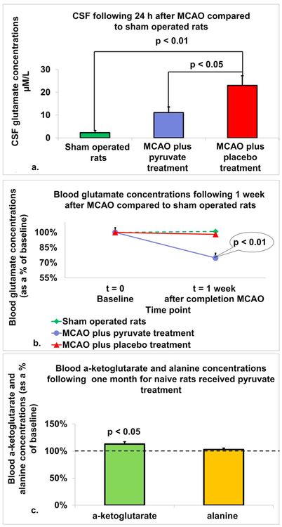
Fig. 3.
In-vivo effects of pyruvate on blood and brain glutamate levels in a rat model of MCAO. a. Concentrations of glutamate in CSF: There was a significant increase in CSF glutamate concentration 24 h post-MCAO in both post-stroke rats treated with placebo and post-stroke rats treated with pyruvate, compared to sham operated rats (p < 0.01). Furthermore, CSF glutamate concentrations were significantly lower in post-stroke rats treated with pyruvate compared to post-stroke rats treated with placebo (p < 0.05). The data are measured in μM/L and presented as mean ± SEM. b. Concentrations of glutamate in blood: There was a significant decrease in concentrations of glutamate in the blood 1-week post-MCAO in rats treated with pyruvate compared to post-stroke rats treated with placebo and sham operated rats (p < 0.01). The data are measured as μM/l and expressed as a percentage of the baseline ± SD. c. Serum blood concentrations of alanine and α-Ketoglutarate: In naïve rats treated with pyruvate for 1 month, concentration of α-ketoglutarate in blood serum was significantly increased compared to baseline levels (p < 0.05). While concentration of alanine in blood serum was increased compared to baseline, the difference was not statistically significant. The data is presented as average percentage of baseline ± SEM.

