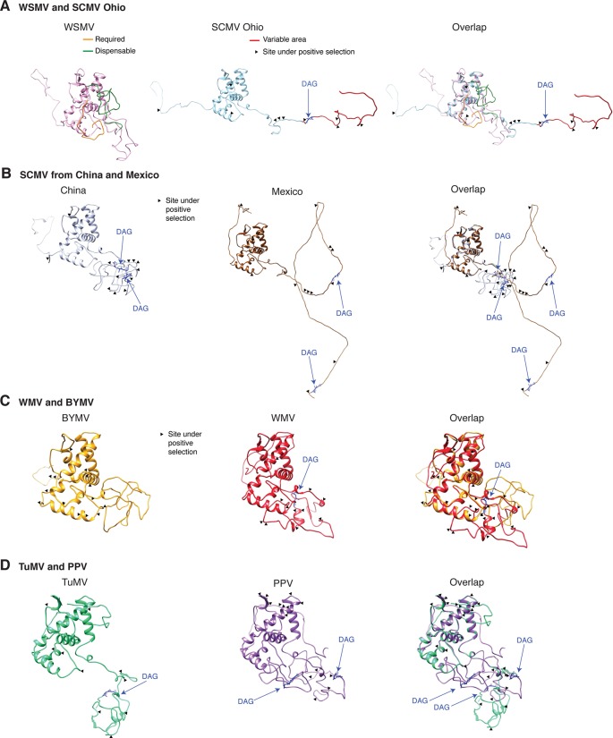
Figure 14.
Schematic representation of the coat protein structural model. For potyviruses with the most amino acid variation, models were generated and superimposed using Phyre2 and Chimera v1.13, respectively. Wheat streak mosaic (WSMV; NC_001886) was used as a control. The coat protein folds into a central core, an N terminal and a C terminal variable loops. TM score and RMSD for each of the analyzed pairs were 0.49 ± 0.60 and 2.0 ± 2.4, respectively. Positive selection sites and the DAG motif are indicated. (A) Model for the Ohio isolate (AFQ35988.1) of SCMV compared to WSMV. (B) Representative SCMV isolates from China (AGE32037.1) and Mexico (ADG23201.1). (C) Bean yellow mosaic virus (NP_612218.1) and watermelon mosaic virus (ABD59007.1). (D) TuMV (NP_062866.2) and PPV (AFJ74692.1).

