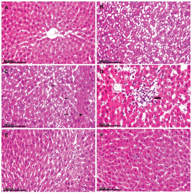
Figure 3.
Photomicrograph of liver tissue sections stained with (H&E) of (A) normal rat showing normal hepatocytes. (B–D): CuO-NPs intoxicated rats showing. (B) Diffuse vacuolization of hepatocellular cytoplasm. (C) Cytoplasmic vacuolization (arrow) in some hepatocellular cells and sporadic necrosis (arrowhead) and abundant apoptosis in others as well as the presence of intracytoplasmic eosinophilic globular inclusions. (D) Focal area of coagulative necrosis infiltrated with mononuclear inflammatory cells (arrow). (E) CuO-NPs + 1 mL/kg bwt PJ group showing mild cytoplasmic vacuolization with few apoptotic bodies. (F) CuO-NPs + 3 mL/kg bwt PJ group showing normal hepatocytes with sparse apoptotic figures.

