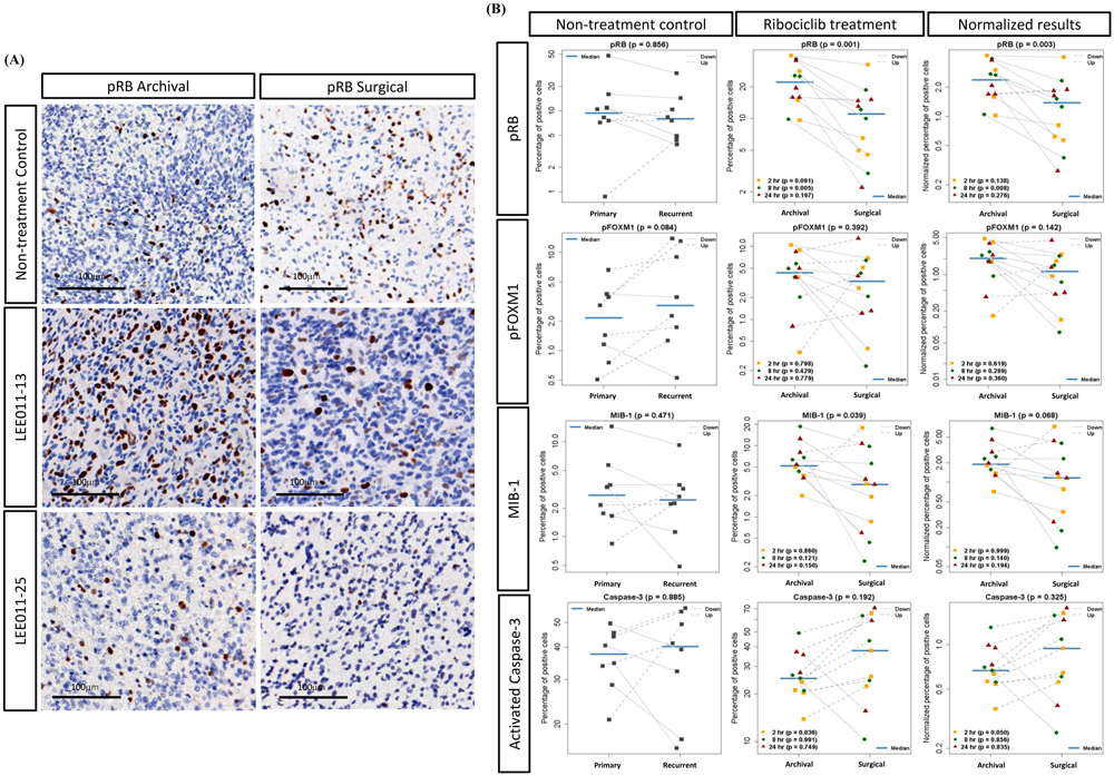
Figure 3.
Pharmacodynamic analysis of surgical glioblastoma tissue after short-term ribociclib treatment. (A) Representative pRB immunohistochemistry staining for matched archival and surgical specimens from ribociclib treatment patients and historical control patients are shown. The scale bar is 100 μm. (B) Quantification of pRB, pFOXM1, MIB-1, and activated caspase-3 positive cells by Aperio imaging system from 8 matched archival and 12 surgical specimens are shown. Three time-escalating cohorts (2-, 8-, and 24-hour) after the last dose of ribociclib are indicated. Non-treatment control samples are in the left column whereas the treatment group results and the normalized results are shown in the middle and right columns. P-value of each marker is indicated.

