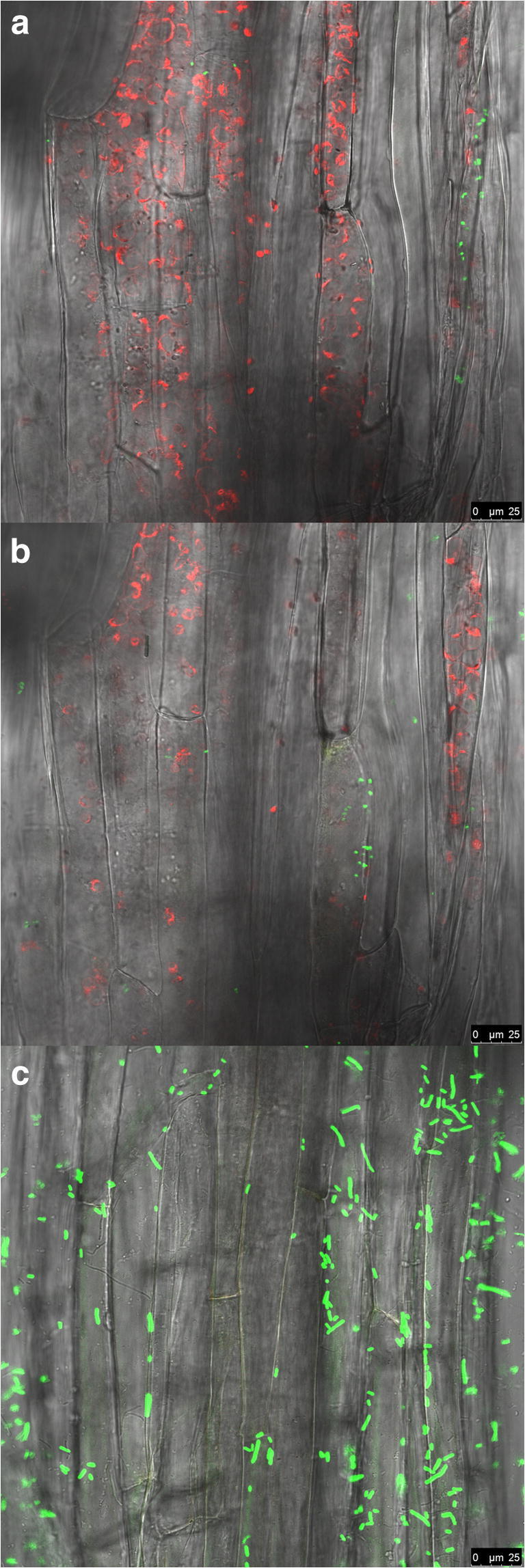
Fig. 2.

Confocal laser scanning microscopy micrographs of gfp/gusA-tagged Serratia sp. M24T3 cells colonizing A. thaliana roots. Longitudinal section of A. thaliana root inoculated with strain M24T3 and grown for 7 days in a 12 multiwell plate. The images a–b show different optical sections through one sample after M24T3 (green) colonizing a inner tissues, b intercellular spaces as well as in the cortex region. Chloroplasts are visible in red fluorescence. c Negative control with gfp/gusA-tagged E. coli (green) outside the tissue (top layer) (bar 25 μm)
