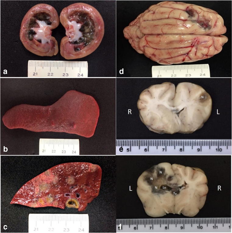
Fig. 1.
Gross findings associated with disseminated phaeohyphomycosis in a mixed-breed dog. Observe the black-green accumulation within the medullary and the white substance at the pelvic regions of the left kidney (a) and the nodule at the spleen (b). There are multifocal to coalescing necrotic regions at the sectioned surface of the liver (c). Observe two green-black areas at the frontal lobe of the cerebrum (d). Transversal sections of the brain demonstrating the extension of the black-green necrotic (N) regions, which extended from the left frontal lobe (e) to the caudal part (f) of the brain (R, right; L, left). There is mild dilation of the left lateral ventricle (Lv) due to the accumulation of necrotic material; necrotic debris was also observed in the third ventricle (Tv). Scale in centimeters

