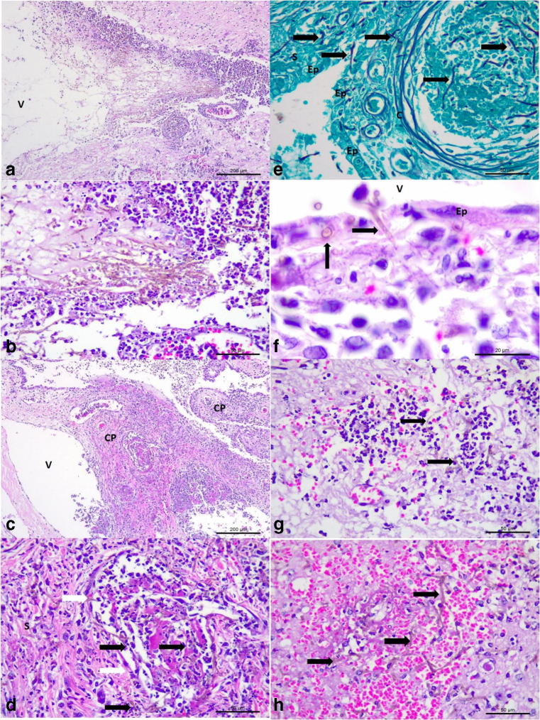
Fig. 3.
Histopathologic and histochemical findings associated with mycotic ventriculitis and choroid plexitis in a mixed-breed dog. Mycotic ventriculitis: observe the dilated lateral ventricle (V) and severe accumulations of pigmented fungal septate hyphae (a); closer view showing the myriad of pigmented hyphae admixed with neutrophils and macrophages (b). Mycotic necrotizing choroid plexitis: there is severe necrosis of the stroma and ependymal cells of the choroid plexus (CP) within the lateral ventricle (V) of the brain (a). Closer view of the choroid plexus demonstrating a mycotic thrombosis at the stroma (S) of the choroid plexus; observe intravascular fungal hyphae (black arrows) within the stroma and a capillary (white arrows) of the choroid plexus (d). There is destruction of the ependymal membrane (Ep) and accumulations of fungal hyphae within a capillary (c) and the stroma of the choroid plexus (e). There are pigmented fungal organisms (black arrows) within the necrotic wall of the ependymal membrane (f), and within a microabscesses (g), and hemorrhage of the cerebrum (h). Hematoxylin and eosin stain, a–d, f–h; Gomori methenamine-silver histochemical stain, e. Bar, a, c, 200 μm; b, d, e, g, and h, 50 μm; f, 20 μm

