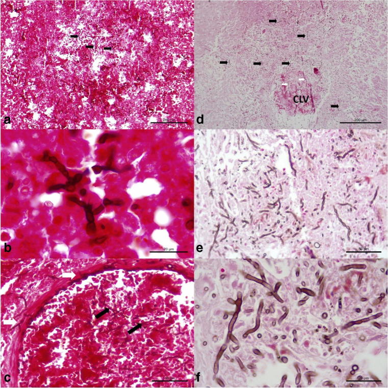
Fig. 5.
Histochemical identification of intralesional septate fungal organisms in a dog with disseminated cladosporiosis. Observe intralesional pigmented fungi (black arrows) in the brain (a, b), and within an artery of the cerebrum (c). There are numerous accumulations of intralesional fungi in the parenchyma (black arrows) and within (white arrows) the affected central lobular vein (CLV) of the liver. Closer approximation (e and f) to demonstrate the intralesional fungal organisms within the liver. Fontana-Masson stain; bar, a, d, 200 μm; b, f, 20 μm; c, e, 50 μm

