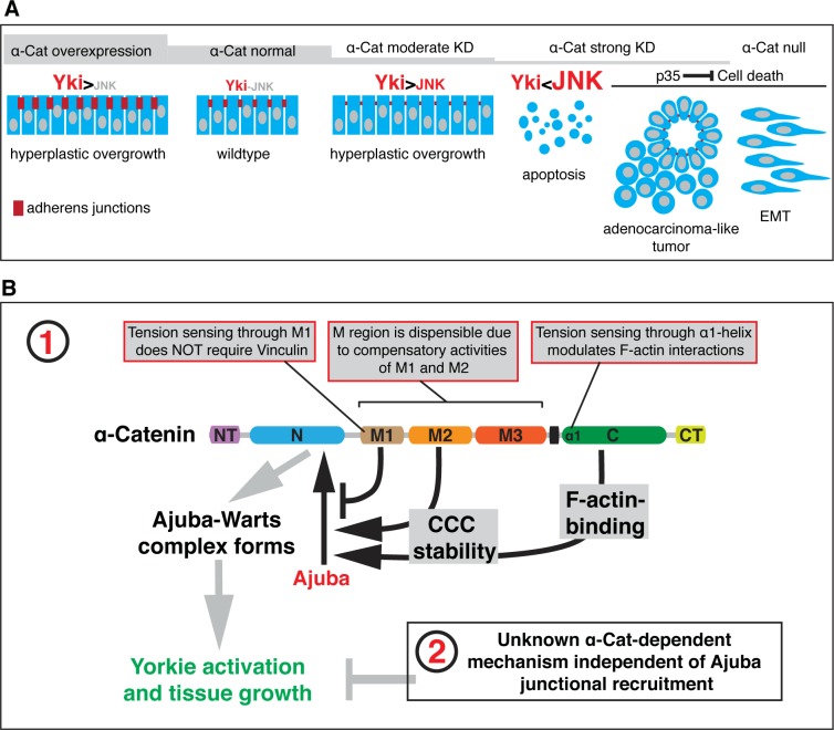
Fig 10. Model for α-Cat function in the regulation of tissue growth.
(A) Illustration of the α-Cat phenotypic series and model for the differential activation of JNK and Yki signaling in response to changes in α-Cat levels (see text for further discussion). (B) Schematic model of α-Cat function in the regulation of tissue growth of the wing disc epithelium. α-Cat regulates tissue growth through Ajuba (Jub)-dependent (1) and Jub-independent (2) mechanisms. M1 limits Jub recruitment to the N domain of α-Cat by a mechanism that is not understood, but does not involved the main known binding partner of M1, Vinc. Loss of M1 compromised M region mechanosensing and led to a high-level constitutive recruitment of Jub that is independent of tissue tension. The M2 domain (and to a lesser extent M3) and the α-Cat ABD regulate stability of the CCC, with lower CCC levels at AJs reducing Jub recruitment. Loss of ABD mechanosensitivity by compromising α1-helix causes increased F-actin binding and stabilizes the CCC, leading to enhanced Jub recruitment and consequently tissue growth.

