
Figure 3:
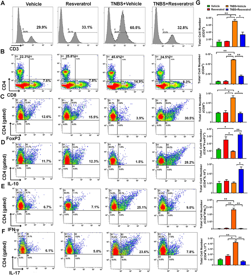
Resveratrol alters T cell subsets during TNBS colitis. The study was designed as described in Fig 1 legend. Flow cytometry histograms/dot plots are shown for the following T cell subsets: CD3+ (A), CD4+ or CD8+ cells (B), CD4+FOXP3+ (C), CD4+IL10+ (D) and CD4+IFNγ+ (E), and CD4+IL-17+ (F) expressing cells. For Figures C-F, cells were gated on the CD4+ population. The gating strategy for the CD4+ populations is detailed in Supplemental Figure 2. Quantitative bar graphs depicting absolute cell numbers of the T cell subsets is provided (G) Each experimental group had at least 5 mice included, and significance (p-value: *<0.05, **<0.01, ***<0.005, ****<0.001) was determined for absolute cell numbers by using one-way ANOVA followed by Tukey’s post-hoc multiple comparisons test. Data is representative of at least 3 independent experiments.