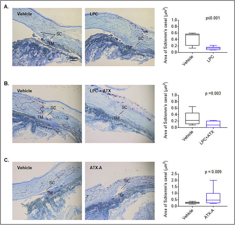
Figure 9: LPC, ATX and ATX inhibitor induced changes in Schlemm’s canal histology in ex-vivo perfused mouse eyes.
Semi-thin plastic sections of perfused mouse eye anterior segments stained with methylene blue and examined under light microscopy revealed that the Schlemm’s canal area in eyes perfused with LPC (3 μM; A) and LPC (3 μM) plus ATX (2 μg recombinant mouse; B) was significantly reduced by ~70% (0.13±0.02 μm2) and ~77% (0.07 ± 0.02 μm2) respectively, compared to eyes perfused with vehicle (0.42±0.06 μm2 and 0.30±0.06 μm2, respectively). In contrast, perfusion of eyes with ATX inhibitor (1 μM ATX-A) resulted in a significant increase in the Schlemm’s canal area by ~129% (0.62±0.13 μm2) relative to vehicle perfused eyes (0.27±0.02 μm2). Each treatment included a paired contralateral eye as a control. For each treatment, two eyes were analyzed and 3 semi-thin sections used per specimen to calculate the area of the Schlemm’s canal tissue using ImageJ software.

