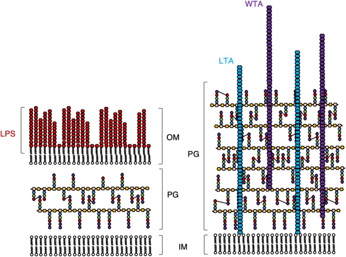
Figure 2.

Comparison of the cell envelopes of Gram‐negative (left) and Gram‐positive (right) bacteria. The cell envelope of Gram‐negative bacteria comprises of an inner (IM) and outer membrane (OM) decorated with lipopolysaccharide (LPS), sandwiching a relatively thin layer of peptidoglycan (PG). By contrast, the cell envelope of Gram‐positive bacteria is comprised of a single cell membrane surrounded much thicker PG layer complemented with lipoteichoic acid (LTA) and wall teichoic acid (WTA). Cross‐links between peptide stems are shown as three to four cross‐links for the sake of simplicity. Not shown are the multitude of proteins that sit in the inner and OM of Gram‐negative bacteria, nor those that reside in the membrane or the PG of Gram‐positive bacteria. The components of PG are displayed using the same scheme as in Figure 1, with the nascent PG chain containing D‐Ala at positions 4 and 5. The mature PG is represented without the D‐Ala at position 5 and occasionally also without the D‐Ala at position 4 to represent the natural variability of the peptide stem in the mature PG mesh
