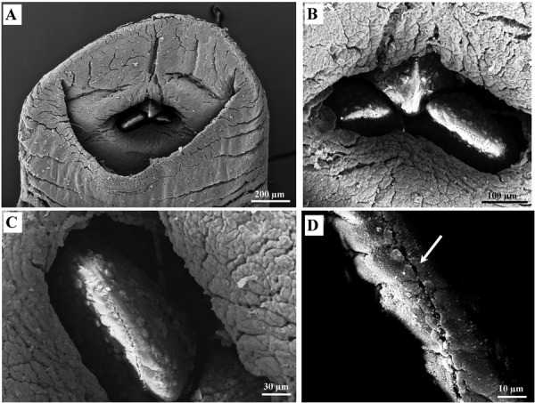
Fig. 4.

Scanning Electron Microscope images of anterior sucker of a Limnatis nilotica specimen. Oral sucker and the three jaws in the bottom (A), The jaws are in form of a triangle (B) bearing a number of rounded papillae on both sides (C) and a line of tooth craters in the apex (white arrow) (D).
