Figure 3.
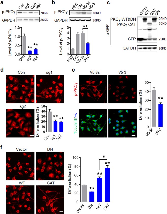
Activation of PKCγ is essential for neuronal differentiation of N2a. (a) Western blot analysis of PKCγ depletion in N2a cells mediated by CRISPR/Cas9. (b) Western blot analysis showed that V5-3 inhibited PKCγ activation induced by 30% OM in N2a. (c) Western blot analysis indicated various PKCγ lentiviral constructions were expressed in N2a. (d) PKCγ depletion inhibited neuronal differentiation induced by 30% OM. N2a were immunostained with Tubulin III antibody and the percent of differentiated N2a cells with GFP+ was quantified. (e) V5-3 (20 nM) attenuated neuronal differentiation of N2a. (f) Activation of PKCγ facilitated neuronal differentiation of N2a. N2a cells were infected with indicated PKCγ constructs and induced differentiation. For d and f, the percent of differentiated cells with GFP+ was quantified in right panel. All data are expressed as mean ± SD (n = 3 individual experiments in each group) and compared by one-way ANOVA (a,b,d,f) or Student’s t-test (e) (**p < 0.01, #p < 0.05 vs indicated group). Scale bars, 20 μm.

