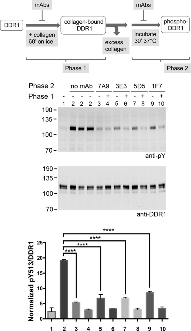
Figure 5.
Anti-DDR1 mAbs block phosphorylation of collagen-bound DDR1. The diagram at the top gives an overview of the experimental procedures. HEK293 cells transiently expressing wide-type DDR1 were first incubated with collagen I for 60 minutes on ice, in the presence (+) or absence (−) of the indicated anti-DDR1 mAbs (Phase 1). Following washes, cells were incubated for 30 minutes at 37 °C, in the absence (no mAb) or presence of the indicated mAbs (Phase 2). The sample shown in lane 1 was lysed immediately after the incubation with collagen on ice. Samples labelled 2 were replicate lysates from 3 different wells. Cell lysates were analysed by Western blot using a mAb against phosphorylated Tyr-513 (anti-pY). The blot was stripped and re-probed with rabbit anti-DDR1. The positions of molecular mass markers are indicated on the left in kDa. The bar chart shows the densitometry analysis of pY513 band intensities after normalization to total DDR1. Each value is a percentage of the sum of all the pY513/DDR1 signals on the blot. The graph shows mean band intensities + SEM (N = 3). ****p < 0.0001 (one-way ANOVA, followed by Dunnett's multiple comparison test).

