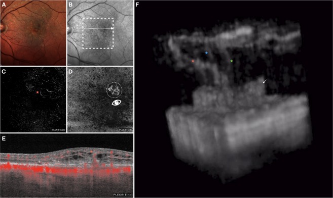
Figure 3.
Multimodal imaging of the LE of an 82-year-old woman diagnosed with type 3 neovascularization. Multicolor (A) and infrared (B) images show areas of RPE alteration and mottling in the macula. En face OCTA images segmented at the outer retina (C) and RPE-RPE fit (D) slabs show one tuft-shaped high-flow lesion (highlighted with an orange asterisk) and presence of a sub-RPE neovascularization, respectively. The white arrow on the infrared reflectance image shows the location and direction of the OCTA B-scan (E), this displaying an high-flow vessel originating from the deep vascular complex and reaching the sub-RPE space. The 3D OCTA visualization (image F is the frame obtained by visualizing the circular region of interest highlighted on image D and visualized from the angle marked with the white eye) revealed the presence of three distinct intraretinal lesions with a filiform shape (marked with 3 asterisks), which merge in the outer retinal layers, and one sub-RPE neovascularization (white arrow).

