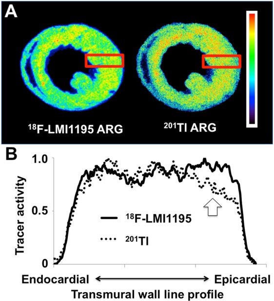
Figure 2.

(A) Representative cross-sectional short-axis images at a midventricular level on dual-radiotracer autoradiography with 18F-LMI1195 and 201Tl. 18F-LMI1195 distribution was homogeneous throughout the left ventricular wall and no significant differences in radiotracer activity were detected in mid-ventricular short axis slices. This is in contradistinction to the autoradiography (ARG) of 201Tl: A slight discrepant uptake pattern in the subepicardial wall portion can be appreciated compared to 18F-LMI1195 ARG. (B) Transmural wall line profile of both 18F-LMI1195 and 201Tl. The macroscopic findings were further corroborated quantitatively: Compared to the subendocardial wall section, radiotracer activity for 201Tl was significantly lower in the subepicardial wall portion. 18F-LMI1195 remained stable over all three section (subendocardial portion, mid-portion, and subepicardial portion).
