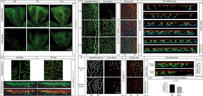
Fig. 2.
Retinal vascular inner plexus develops coaxially to the overarching veins. a Morphology of the superficial (upper) and inner (lower) vascular plexus from Cldn5-GFP transgenic reporter mouse retina at P8, P9, and P10. The vascular network is genetically labeled (GFP, shown in green). “A” and “V” indicate the position of an artery and a vein, respectively. b Visualization of the superficial vascular layer around an artery (left) and a vein (right) of Cldn5-GFP mouse retina at P8. The EC projections toward the inner plexus specific to each area were reconstructed (3D vertical view). Retinal EC were genetically labeled by Cldn5-GFP (green) and further stained for ICAM2 (red) and phospho-histone H3 (pHH3, gray) to visualize vascular lumen and proliferation, respectively. Depth coding of the same area is shown. c Superficial (left), inner (middle left) vascular plexus, and 3D overlay view from top (middle right) of Cldn5-GFP mouse retina at P8, P9, and P10. 3D vertical view below the artery or the vein is shown (right). d Superficial (left), inner (middle left) vascular plexus, and 3D top view (middle right) of WT and Rac1 ECKO retina. A vertical view below the vein (V) is shown (right). Artery (A) and vein (V) are indicated. e Quantification of the EC projections to the inner plexus observed below the vein in Rac1 ECKO and WT mouse retina at P8

