
Fig. 6.
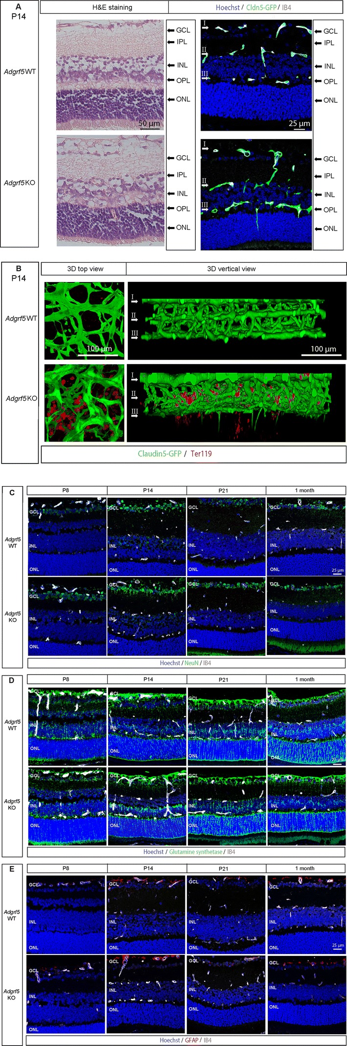
Adgrf5 KO retinal tissue recovers after vessel normalization. a Images from WT and Adgrf5 KO mouse retina at P14. Cross-sectioned retinae were stained with H&E (top) and Hoechst (blue). The vasculature is depicted by IB4 staining (gray) and Claudin5-GFP expression (green). b 3D reconstruction images of the three vascular layers of Adgrf5 KO retina at P14. The retinal structure is presented viewed from the superficial layer (3D top view, left), or from a vertical view (right). Erythrocytes are visualized by Ter119 staining. c Images from WT and Adgrf5 KO mouse retina at P8, P14, P21, and 1 month. Cross-sectioned retinae were stained with anti-NeuN antibody (green), Hoechst (blue), and IB4 (gray). d Images from WT and Adgrf5 KO mouse retina at P8, P14, P21, and 1 month. Cross-sectioned retinae were stained with anti-Glutamine synthetase antibody (green), Hoechst (blue), and IB4 (gray). e Images from WT and Adgrf5 KO mouse retina at P8, P14, P21, and 1 month. Cross-sectioned retinae were stained with anti-GFAP antibody (red), Hoechst (blue), and IB4 (gray)