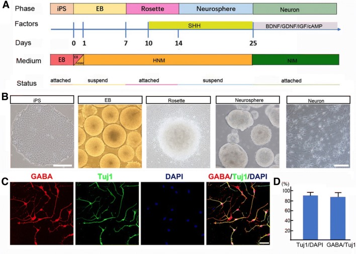
Fig. 5.
Differentiation of iPSCs from an HD patient into ventral GABAergic neurons. A Steps of differentiation of iPSCs. B Typical cellular morphology at the embryoid body (EB), rosette, neurosphere, and neuron stages. Scale bars, 500 µm (iPSC stage) and 200 µm (EB, Rosette, Neurosphere and Neuron stages). C Representative co-immunostaining images of cells stained with Tuj1 and GABA antibodies. Scale bar, 50 µm. D Counts of Tuj1-positive neurons or Tuj1 and GABA double-positive neurons (mean ± SD).

