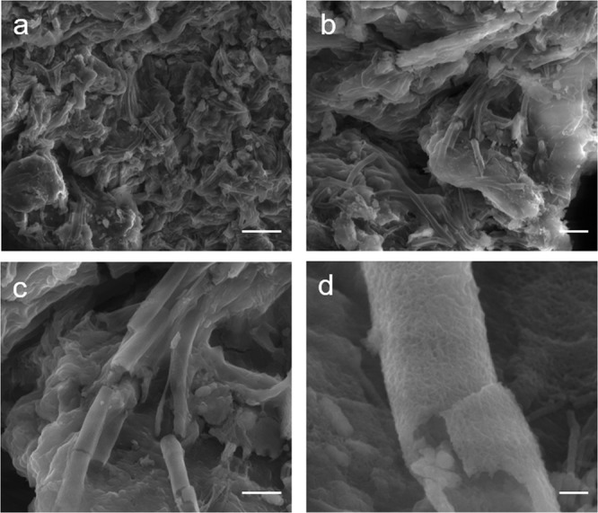
Figure 2.

Structure and morphology of a L. ochracea microcolony cluster on a sediment particle. Scanning electron micrographs using secondary electrons illustrating freshly collected and untreated L. ochracea, collected by D. Emerson. (a) Large cluster of sheaths on a sediment particle. Scale = 10 µm. (b) Cluster of L. ochracea growth. Scale = 5 µm. (c) Detail of L. ochracea cluster showing unidirectional growth pattern. Note the relatively smooth surface texture. Scale = 2.5 µm. (d) Detail of broken L. ochracea sheath. Scale = 0.25 µm.
