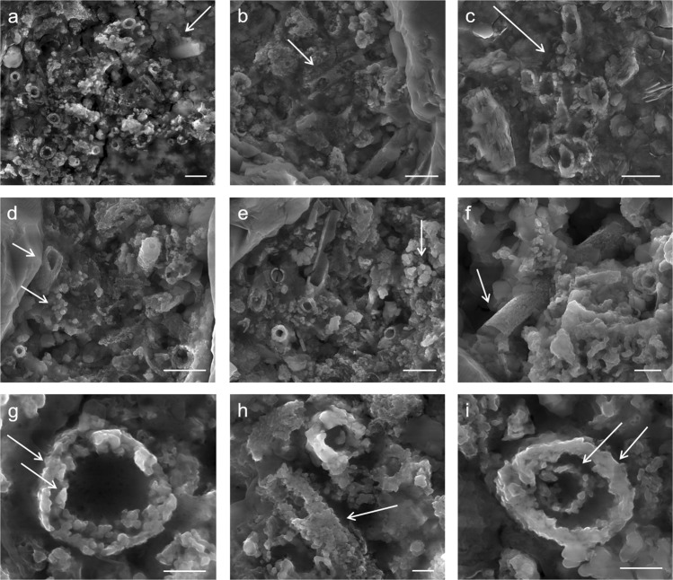
Figure 3.
Selected micrographs from SEM examination of targeted red pigment particle in GcSi-1 rock art sample. (a) Concentration of broken and randomly oriented L. ochracea sheaths. Note the fractured diatom fragment in the upper right corner, and hematite and silicate microspheres distributed throughout. Scale = 2.5 µm. (b) Detail of L. ochracea sheath showing advanced degradation (holes) in the sidewall of a sheath body. Scale = 2.5 µm. (c) Detail of a small, intact cluster of L. ochracea bodies. This was the sole identifiable example of a coherent cluster in the sample. Note the warped shapes of the tube openings. Scale = 2.5 µm. (d,e) Concentrations of L. ochracea sheaths displaying fractured ends and globular, porous sheath exterior walls. Note the clusters of hematite and silicate microspheres and blocky octahedral and tetrahedral hematite polymorphs distributed through the pigment matrix. Scale = 2.5 µm. (f) Rare example of a relatively intact L. ochracea sheath. Scale = 1.25 µm (g–i) Details of broken sheath ends. Note the globular surface texture indicating localized melt and recrystallization of iron and silica. Specimens shown in (g,i) have double-walled inner rings, indicating maturity at the time of death. Scales = 0.5 µm.

