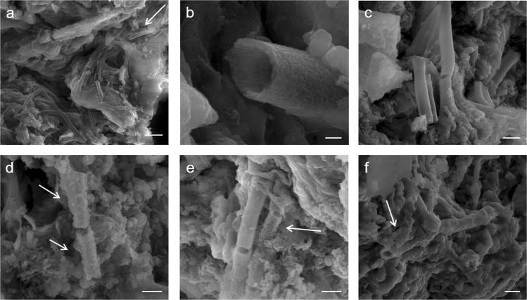
Figure 7.
SEM Micrographs of Incrementally Heat-Treated L. ochracea Control Samples. (a) Untreated FeOB. Note the smooth exterior texture. Scale = 10 µm (b) 200 °C, note the absence of structural changes. Scale = 0.25 µm (c) 400 °C, note the absence of structural changes and smooth sheath exterior. Scale = 1.25 µm (d) 600 °C, showing structural changes including globular deposits and fraying of sheath exteriors. Scale = 0.75 µm. (e) 800 °C, note melt features on sheath exterior surfaces and nucleation of hematite microspheres. Scale = 1.0 µm (f) 1,000 °C, note the complete phase transformation to magnetite and hematite polymorphs. Scale = 1.25 µm. *An early iteration this figure was published in a non-peer-reviewed conference proceedings abstract in a special issue of the journal Microanalysis and Microscopy87.

