Figure 1.
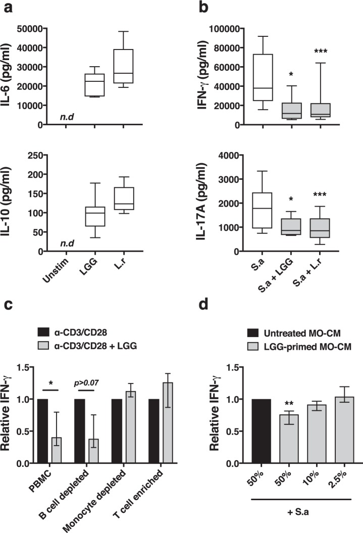
Lactobacilli-CFS dampens IFN-γ in a monocyte-dependent manner. PBMC were stimulated as indicated and culture supernatants were collected after 48 h and secreted cytokines evaluated with ELISA. (a) Secretion of IL-6 and IL-10 following stimulation with LGG- or L. reuteri (L.r)-CFS (n = 5–6). (b) Secretion of IL-17A and IFN-γ following stimulation with S. aureus (S.a)-CFS alone and in combination with Lactobacillus-CFS (n = 6–14). (c) Whole PBMC, B cell depleted, monocyte depleted or enriched T cells were stimulated with T cell specific activator beads towards CD3/CD28 alone and in combination with LGG-CFS. Secreted levels of IFN-γ was determined and normalized to stimulated cells in the absence of LGG-CFS, (n = 4–8). (d) Monocyte-conditioned medium (MO-CM) from isolated monocytes primed with LGG-CFS for 6 h, extensively washed and incubated in fresh cell culture medium for 14 h, was mixed 1:1 (50%), 1:10 (10%) or 1:40 (2.5%) with S.a-CFS-stimulated autologous PBMC cultures. Secreted levels of IFN-γ was quantified and normalized to stimulated cells mixed 1:1 with unprimed monocyte conditioned medium, (n = 6–8). Boxes cover data between the 25th and the 75th percentile with medians as the central line and whiskers showing min-to-max. Bar plots show median with interquartile range.

