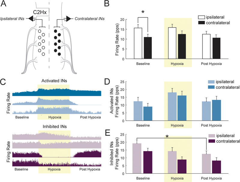
Fig. 3.
A. A schematic illustrating the method for multi-electrode array recordings of spinal INs following C2Hx. B. Average firing rate (pulses per second) of all ipsilateral and contralateral spinal INs before, during (shaded yellow), and after hypoxia. C. Representative compressed firing rate traces, from one experiment illustrating hypoxia-activated neurons (i.e. those which increase firing during hypoxia) and hypoxia-inhibited INs (i.e. those which decrease firing during hypoxia). D. Averaged firing rate of activated and E. inhibited INs before, during (shaded yellow), and after hypoxia. Post-hoc analysis indicated ipsilateral INs had a lower burst rate during the post-hypoxic period as compared to baseline (P = 0.0195, indicated by *).

