
Figure 2: Adult mouse cardiomyocytes can be cultured in 3D PEG hydrogels for extended times.
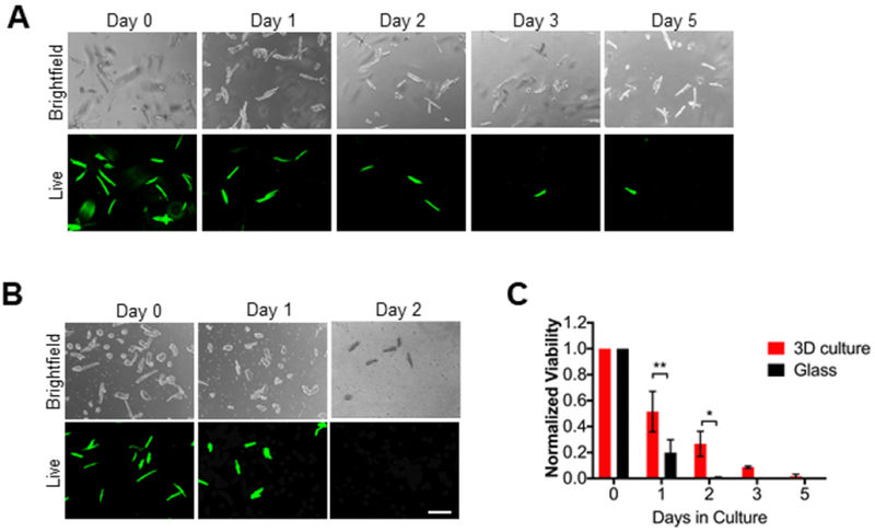
A) Representative brightfield and live images of adult mouse cardiac myocytes (AMVMs) cultured in 3D PEG hydrogels for 0, 1, 2, 3, or 5 days. Cells were cultured with calcein (Live) and ethidium homodimer for 1 hour prior to imaging. Scale bar = 200um. B) Representative brightfield and live images of AMVMs cultured on glass coverslips for 0, 1, 2 days. Cells were cultured with calcein and ethidium homodimer for 30 minutes prior to imaging. Scale bar = 200 um. C) Quantification of AMVM viability in 3D culture and glass coverlsips over 0, 1, 2, 3, or 5 days. For 3D culture N=3 mice, for glass N=4 mice. Two-way ANOVA with Bonferroni post-hoc test applied; *p<0.5 **p < 0.01. Data reported as mean ± SEM.