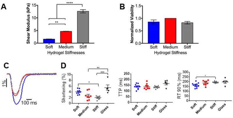
Figure 4: Adult mouse cardiomyocytes contractility is affected by different stiffnesses.

A) Columns showing the shear modulus for different thiol-ene PEG hydrogels, measured by shear rheology. Data reported as mean ± SEM. One-way ANOVA with Bonferroni post-hoc test applied; **p < 0.01, ****p < 0.0001. B) Viability of adult mouse cardiomyocytes encapsulated in thiol-ene PEG hydrogels with different stiffnesses at day 0 (2 hours post-encapsulation). Viability assessed by calcein/ethidium homodimer staining and reported as % normalized to cells encapsulated in medium stiffness PEG hydrogel. Data reported as mean ± SEM, N=3. C) Representative cell shortening traces of adult mouse cardiomyocytes encapsulated in soft, medium, or stiff PEG hydrogels (blue, red, and grey, respectively). D) Graphs showing shortening, time-to-peak (TTP) and relaxation 90% (RT90%) of adult mouse cardiomyocytes encapsulated in soft, medium, stiff PEG hydrogels, or not encapsulated. Data reported as scatter plots with the central line representing the mean value, and the error bars representing SEM from n=10 cells (soft), n=8 cells (medium), n=7 cells (stiff), and n=5 cells (no gel). N=3 (number of animals). One-way ANOVA with Bonferroni post-hoc test applied; *p<0.5 **p < 0.01, ***p < 0.001.
