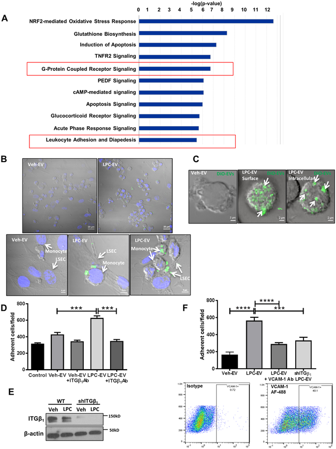
Fig. 3. toxic hepatocyte-derived EVs promote monocyte adhesion to LSECs via an ITGβ1-dependent mechanism.
Lipo (A) Top represented canonical pathways in monocytes stimulated with LPC-EVs vs Veh-EVs. (B) Equal number of Huh7 cells were treated with either veh or LPC. EVs were collected from the conditioned media and labelled with DiO. THP1 cells were co-cultured with human LSECs in the presence of labelled EVs. Scale bar: 20 μm for the top panel, and 5 μm for the bottom panel. (C) Z-stack confocal microscopy of THP1 incubated with DiO-labelled EVs from LPC-treated Huh7 cells (white arrows). (D) Primary mouse monocytes were stimulated with Veh-EV or LPC-EV from PMH ± ITGβ1Ab, and infused in microfluidic chambers coated with a monolayer of primary mouse LSECs. Adherent cells were quantified. (E) Immunoblot analysis showing ITGβ1 knockdown in shITGβ1 cell line. Beta-actin was used as a loading control. (F) THP1 cells were stimulated with either Veh-EV or LPC-EV from wild-type (WT) Huh7 cells, or shITGβ1 Huh7 cells, and infused in microfluidic chambers coated with a monolayer of primary human LSECs ± VCAM-1 Ab. Adherent THP1 cells were quantified similar to D. VCAM-1 is expressed on human LSECs under basal condition as shown by flow cytometry; bar graphs represent mean±SEM; n=6, ***p<0.001, ****p<0.0001 (One-way ANOVA with Bonferroni’s multiple comparison).

