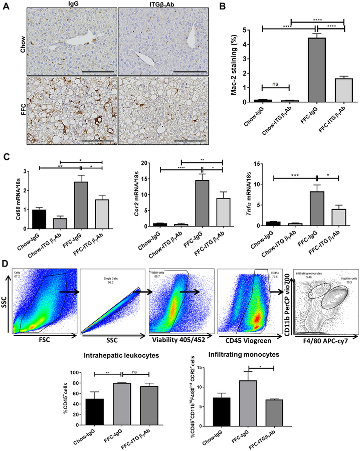
Fig. 5. Anti-ITGβ1 antibody treatment in FFC-fed mice attenuates hepatic inflammation.
(A) Representative images of macrophage galactose-specific lectin (Mac-2) staining of liver sections. (B) Mac-2 positive areas were quantified in 10 random 20x microscopic fields and averaged for each animal. (C) Hepatic mRNA expression levels of Cd68, Ccr2 and Tnf-α were assessed by real-time PCR. Fold change was determined after normalization to 18s expression and expressed relative to Chow-IgG mice. (D) Flow cytometric analysis of the IHL population: top panels show the gating strategy; infiltrating monocytes were defined as CD45+ CD11bhi F4/80int CCR2+cells. Bottom panels show quantification of each population. Bar graphs represent mean±SEM; n=3–5 per group; *p<0.05, **p<0.01, ***p < 0.001, ****p<0.0001 (One-way ANOVA with Bonferroni’s multiple comparison, unpaired t test for panel D.).

