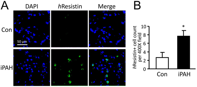
Figure 1.
Expression of hResistin in lungs from patients with iPAH. (A) Paraffin-embedded sections of lungs from normal subjects (Con, n = 6) and patients with iPAH (n = 6) were stained with anti-hResistin Abs (green) and counter-stained with DAPI (blue). Immune cells producing hResistin were observed in iPAH lungs but were infrequent in control tissues. Magnification: 400X. (B) hResistin-expressing cells in the lungs were quantified. Positive cells were counted on 10 randomly chosen fields of lung sections in each patient at 400-fold magnification. Data are presented as means ± SEM, *p < 0.05. Immunofluorescence experiments and analyses were repeated twice.

