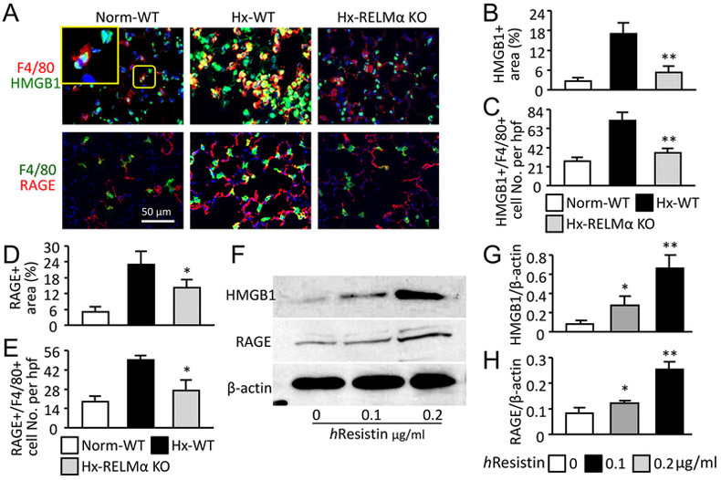
Figure 2.
RELMα/hResistin activates HMGB1/RAGE axis in pulmonary macrophages in vivo and in vitro. (A) Double-color immunofluorescence analysis of lung tissues after 4 days of hypoxia (Hx). Sections were stained with anti-F4/80 Abs and co-stained with anti-HMGB1 or anti-RAGE Abs. The high-magnification inset depicts HMGB1 nuclear localization in the normoxic wild-type (WT) mouse lung. Photographs shown are representative of three independent experiments with four animals per group. Magnification: 400X. Norm, Normoxia; KO, knockout. (B-E) Quantitative analysis of data in (A). Percentage of areas positive for HMGB1 (B) or RAGE (D) in wound sites was determined with Adobe Photoshop software. The F4/80+ pulmonary macrophages expressing HMGB1 (C) or RAGE (E) were also counted and expressed as numbers per high-power field (hpf). Data are presented as means ± SEM (n = 4 animals). *p < 0.05 vs Hx WT mice. (F) Western blot analysis of HMGB1/RAGE expression in the hResistin-treated macrophages differentiated from THP-1 monocytes. (G, H) Quantitative analysis of data in (F). The ratios of HMGB1 (G) and RAGE (H) to β-actin were determined. Data are presented as means ± SEM (n = 6). *p < 0.05, ** p < 0.01 vs control vehicle treatment. Data are representative of at least three independent experiments.

