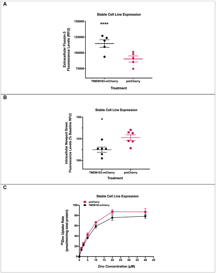
Fig. 1. Functional expression of TMEM163 stably expressed in HeLa cells.
A) Cell membrane impermeant Fluozin-3 (MI-FZ3) fluorescence analysis revealed a significant increase in extracellular RFU levels in the milieu of cells expressing TMEM163-mCherry upon exposure to zinc. Data are represented as mean ± SEM (****p < 0.0001, Student’s t-test, unpaired, two-tailed; n = 5 independent trials). B) Cell membrane permeant Newport Green (MP-NG) fluorescence analysis revealed a significant decrease in intracellular RFU levels of cells expressing TMEM163-mCherry upon exposure to zinc when compared with cells expressing the pmCherry vector control. Data are represented as mean ± SEM of each kinetic time point analyzed (*p = 0.01; Student’s t-test, unpaired, two-tailed; n = 6 independent trials). C) Concentration–d-dependent and saturable uptake of 65Zn by TMEM163-and mCherry-expressing cells. The presence of TMEM163 resulted in significant reduction of intracellular 65Zn levels (p = 0.01; Student’s t-test, paired, two-tailed; n = 3 independent trials).

