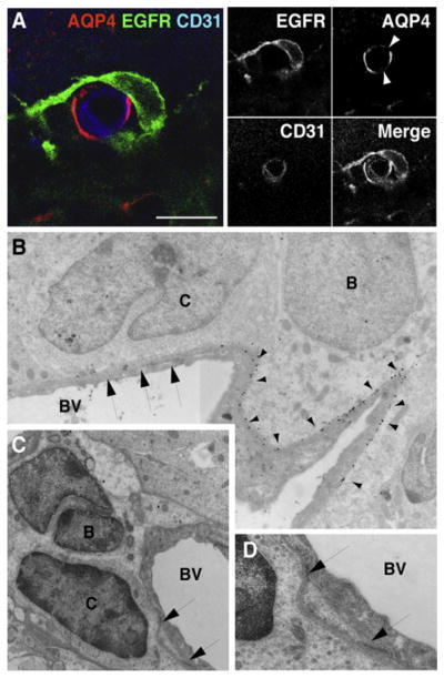
Figure 5. EGFR+ Transit-Amplifying C Cells Contact the Vasculature at Regions of Blood Vessels Lacking Astrocyte Endfeet.
(A) Confocal images of SVZ blood vessels in cross-section showing transit-amplifying C cells (green) contacting the vasculature (blue) at AQP4-negative (red) patches (arrowheads) on blood vessels. Serial optical sections of cell in (A) are shown in Figure S7. Scale bar, 10 μm.
(B) Composite electron micrograph depicting contact between transit-amplifying C cell (labeled C) and blood vessel (BV) at a region of the vasculature that lacks AQP4 staining (arrows). Note the gold particles from AQP4 labeling (arrowheads) on the SVZ astrocyte (type B cell). Magnification, 15,800 ×.
(C and D) Electron micrographs showing an example of a transit-amplifying C cell (labeled C) directly contacting a blood vessel (BV). Image in (D) depicts high-power view of contact points shown in (C). A low-power view of this cell is shown in Figure S8. Magnification, (C) 15,800 ×; (D) 52,900 ×.

