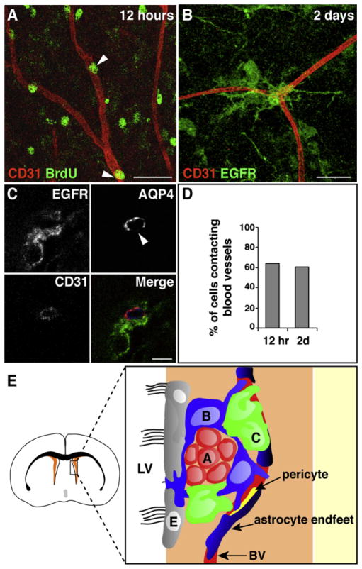
Figure 7. SVZ Stem Cells and Transit-Amplifying Cells Contact Blood Vessels during Regeneration.
(A) At 12 hr after cessation of Ara-C treatment, BrdU+ cells in the SVZ (green, arrowheads) are immediately adjacent to the vasculature (red). Scale bar, 30 μm.
(B) After 2 days, EGFR+ C cells (green) contact blood vessels (red). Scale bar, 30 μm.
(C) Regeneration (EGFR+ cells, green) occurs at sites on blood vessels (blue) that lack AQP4 staining (red). Scale bar, 5 μm.
(D) Histogram showing percentage of cells contacting blood vessels at 12 hr and 2 days after cessation of Ara-C treatment.
(E) Model of the vascular SVZ stem cell niche. Schema of a coronal brain section, with SVZ in orange. Box is expanded to the right to show SVZ cells. Blood vessels (BV) are an integral component of the SVZ niche. Stem cell astrocytes (labeled B, blue) and transit-amplifying C cells (C, green) often contact the vasculature at regions on blood vessels lacking astrocyte endfeet (dark blue) and pericyte coverage (yellow), giving them direct access to vascular and blood-derived signals. Stem cell astrocytes also contact the lateral ventricle. Chains of neuroblasts (labeled A, red) are less closely associated with the vasculature. Ependymal cells (E, gray) line the lateral ventricles.

