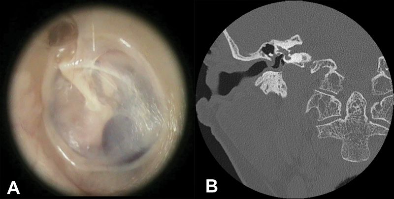
Fig. 2.

Panel A : Left ear. Endoscopic view. A high positioned and dehiscent jugular bulb in contact with the tympanic membrane at the posteroinferior quadrant of the middle ear cavity; Panel B : Computed tomography scan. Coronal view. Right side. Presence of high positioned and dehiscent jugular bulb partially covering the round window niche.
